Anomaly Scan: An Effective Tool To Detect Defects In The Baby Before Birth
4 Dec 2017 | 5 min Read
Babychakra
Author | 1369 Articles
An anomaly scan detects birth defects in babies during pregnancy
Anomaly scan: what is it?
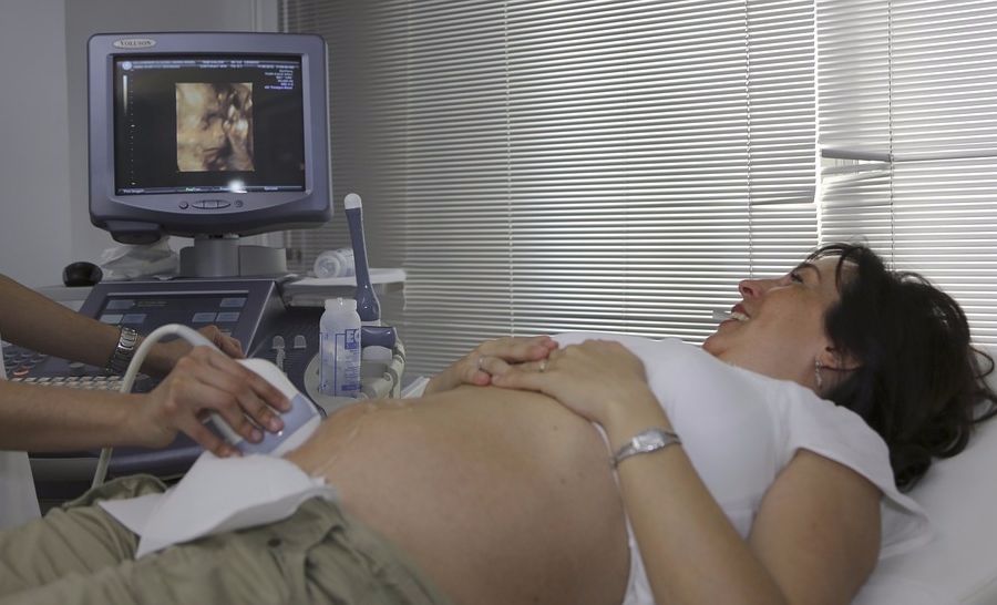
Anomaly scan, also known as fetal anomaly scan or level 2 anomaly scan, or targeted imaging for fetal anomalies, is an ultrasound scan that is done in the second trimester of pregnancy. It is commonly done between 18 to 23 weeks of pregnancy to evaluate the development and growth of the baby. The formation of the baby’s organs is complete by the time pregnancy reaches the second trimester. During this period, all organs are easily visualized in the anomaly scan of fetus.
Why do an anomaly scan?
It is essential to do an anomaly scan of your baby before it is born, to identify any birth defects that may be present and take appropriate measures if required.
The anomaly scan can detect major defects like the following:
- Spina bifida (open spinal cord)
- Anencephaly (defect in the baby’s head), skull
- Abnormality of hands and legs
- Defects in organs like abdomen, kidneys, heart (congenital heart defects), etc.
- Defects of diaphragm (hernia of diaphragm), a muscle that separates the chest and abdomen
- Collection of excess fluid in the brain (hydrocephalus)
- Defects in the face and mouth (cleft lip)

Source: cdc.gov
- Certain genetic anomalies like autism, cerebral palsy, etc. are not detected on the anomaly scan in pregnancy.
- Another chromosomal/genetic birth defect called Down’s syndrome is commonly associated with defects in the heart and abdomen. However, the diagnosis is confirmed only after performing other special genetic tests like amniocentesis, chorionic villus sampling, and non-invasive prenatal testing.
Apart from identifying physical defects in the developing baby, an anomaly scan report also remarks on following aspects:
1. Placenta
- The placenta can be located on the front wall of the uterus (anteriorly placed) or on the backside wall (posteriorly placed), or at the top of the uterus (fundus).
- It may be low lying, covering the neck of the uterus. In such a case, you will be asked to repeat the anomaly scan after 20 weeks or in the third trimester when the placenta changes location with the movements of the growing baby.
2. Gender of the baby.
- The sex of the fetus can be identified using an anomaly scan. However, since preference to a boy over a girl child in India has led to increase in illegal abortions, revealing the gender of the unborn baby is a punishable crime by law. Anomaly scan is not to be used for sex determination of the unborn baby.
3. Umbilical Cord
- The scan looks for knots and total number of blood vessels in the cord.
4. Amniotic Fluid
- The volume of amniotic fluid in which the baby floats plays an important role in evaluating the growth of the baby and delivery date.
5. EDD (Expected Date of Delivery)
- The scan also calculates the age of the baby depending on the growth, development and maturation of various organs. Based on this, the scan helps in giving the expected date of delivery or child birth.
Anomaly scan format
Normally, the anomaly scan has predefined guidelines set for the measurement of certain factors in the growing baby. The baby’s measurements should correspond to the age set in the guidelines. The factors include:
- Head circumference (HC)
- Bi Parietal diameter (BPD)
- Abdominal circumference (AC)
- Femur length (FL)
Ultrasound soft markers
There are certain variants or small deviations in normal findings of the 2 trimester anomaly scan. When present singly or not to a very significant degree, these findings do not point to any abnormality in the unborn baby. These are called as ultrasound soft markers.
Single or very slight deviations are very commonly seen in a normal baby and are not considered abnormal.
When more than one or two prominent soft markers are seen on the scan, it increases the doubt of chromosomal defects.
Ultrasound soft markers taken into account while diagnosing a genetic abnormality include:
- Nuchal fold thickening (fluid collection on the back of the baby’s neck)
- Choroid plexus cyst (cyst in the brain)
- Renal pelvic dilatation (abnormality in the kidney)
- Echogenic foci (collection of amniotic fluid in the abdomen or near the heart)
- Single artery in the umbilical cord– normally umbilical cord consists of two arteries and one vein.
- Hydrocephalus (fluid collection in the brain)
Under normal conditions, ultrasound soft markers are usually present temporarily and disappear in the subsequent anomaly scan at 33 weeks.
Anomaly scan of a genetic defect, like Down’s syndrome shows the presence of nuchal fold, not well formed nasal bones, short hands and legs, curved little finger, renal pelvic dilatation, etc.

Disclaimer: The information in the article is not intended or implied to be a substitute for professional medical advice, diagnosis or treatment. Always seek the advice of your doctor.
Also Read – All You Needed to Know About The Transvaginal Scan (TVS)
A


Related Topics for you
Suggestions offered by doctors on BabyChakra are of advisory nature i.e., for educational and informational purposes only. Content posted on, created for, or compiled by BabyChakra is not intended or designed to replace your doctor's independent judgment about any symptom, condition, or the appropriateness or risks of a procedure or treatment for a given person.
